N-Form® Multi-Electrode Array
Plexon Inc and Modular Bionics™ Inc. have partnered to offer the N-Form®, a customizable 3D multi-electrode array for chronic in vivo electrophysiology. The N-Form array combines features of the Utah array and linear arrays, such as the Plexon V-Probe, for chronic, 3D volumetric recordings. Each array is composed of a grid of linear arrays, or shanks, that are continuous with the backplate. The N-form thus offers the advantage of all components being one monolithic piece, rather than multiple pieces assembled together like other multi-electrode arrays such as the Utah array.
The customizable N-Form® design allows for targeting large volumes of neurons across layers and columns of cortex. The N-Form® can be used as a chronically implanted electrode floating interface allows for long-term, chronic recordings. Stable recordings have been demonstrated for over 9 months in marmosets and cats, with the potential for much longer recordings (longevity study ongoing). Each N-Form® array has a hook on the backplate that enables an insertion tool to be attached to aid with placement in the brain.
The N-Form has already been adopted by researchers at organizations such as University of Pittsburgh, University of Chicago, Brown University, NIH, Northwestern University, Arizona State University, and more.


The next generation of multi-electrode array: multichannel neural recording
 Over the past two decades, researchers using arrays like the Utah array have faced limitations in recording large populations of neurons, including an inability to reach deep brain structures and low-dimensional recordings. Neurons aren’t exclusively arranged laterally on a 2D space, so why should that be all an electrode array can cover?
We teamed up with Modular Bionics to address these limitations with the N-Form array. The N-Form is a customizable 3D array exclusively offered by Plexon. With the N-Form, neuroscientists have the power to choose from a number of recording sites in a three-dimensional space. The animation here demonstrates the N-Form with 32 recording sites at varying depths across four shanks up to 3.5mm in length. Arrays are currently available for up to 128 channels with a maximum length of 15mm. It is compatible with Plexon and non-Plexon systems, and is priced similar to standard linear arrays. Real data from chronic recording experiments and other useful information can be viewed under the Resources tab.
Discuss your needs with a Plexon Sales Engineer to determine the best configuration for your upcoming experiments. It may also be possible to produce arrays beyond the specifications outlined in the Technical Specifications tab.
Features & Benefits
- Multichannel configuration
- Up to 128 channels can be defined within a three-dimensional space
- Customizable designs in terms of length and location of recording sites
- High-quality neural recordings accessible immediately after implantation
- Floating neural interface
- Sterilizable
- Excellent stability in acute and chronic applications
- Microstimulation-capable
- Various connector options are available
Research Applications
In the past year, many labs across several institutions have adopted the N-Form. Their research includes:
- Electrical recording and stimulation of:
- Motor cortex
- Sensory cortex
- Spinal cord
- Peripheral nerve fibers
- and more..
What makes the N-Form superior to the Utah array?
1. Utah Array: limited to 1.5mm in length. N-Form: up to 15 mm.
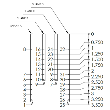
2. Utah Array: limited to one recording site per shank. N-Form: allows you to define specific recording locations at multiple points on each shank (see graphics below)
Utah: Forced to record from only one site for each shank. Two-dimensional:
N-Form: You define the location of electrode sites on each probe. Three-dimensional, with ability to have 8 sites on each shank:
Example above demonstrates N-Form with 32 recording sites at varying depths across four shanks up to 3.5 mm in length
The illustration below depicts the anatomy of a 3.5 mm 4-shank N-Form® Array.

N-Form® Arrays are customizable, as can be seen from the table below. It may also be possible to produce arrays beyond the specifications below. As a result, it is recommended that you discuss your needs with a Plexon Sales Engineer to determine the best configuration and if your unique request can be accommodated.
Technical Specifications
| Electrode diameter | 25 μm, circular cross-section | |
| Electrode material | platinum/iridium | |
| Shank thickness | 125 μm* | |
| Distance between shanks | GEN1: 400 μm GEN2: 500 μm | |
| Maximum number of channels/shank | 8 | |
| Sterilization Method | EtO | |
| Implantable | Yes, insertion holder required for chronic implantation | |
| Electrode Coating | iridium oxide |
Customizable Options
| Cable length | 1.5 cm – 13.0 cm | ||
| Shank length | 500 μm – 15 mm | ||
| Site location | All shanks must have an electrode site at the tip. The site neighboring the tip site must be at least 250 μm away from the tip site. 125 μm spacing between other sites.Shanks >4.00 mm cannot have sites placed more proximal than 3.0 mm from the tip.Shanks between 3.625 mm and 3.875 mm cannot have sites placed more proximal than 2.5 mm from the tip. | ||
| Number of shanks | 1-16 | ||
| Channel count | 4-128 | ||
| Connector Type | Omnetics 32m-V, Omnetics 16m-V, TDT ZC32 , Samtec FTSH-118-01-L-DV, CON/64f-Samtec: SEAF8-10-05.0-S-08-2-k, and CON/128f-Samtec: ADF6-20-03.5-L-04-2 | ||
| Backplate thickness | GEN 1 < 3.5 mm = ~500 μm GEN 1 > 3.5 mm = ~800 μm GEN 2 < 3.5 mm = ~600 μm GEN 2 > 3.5 mm = ~900 μm Single shanks available without a backplate | ||
*Longer shanks may require greater thickness near the base of the shank
N-Form® Arrays are manufactured by Modular Bionics ™ Inc.
Connectors: Omnetics, Zif, and Samtec


Simple Insertion
The insertion tool is easily adaptable to many types of microdrives / inserters. Inserting your high-density microelectrode array into brain, spinal-cord and peripheral-nerve tissue is a high-speed, straightforward procedure.
2025
2024
2023
2022
2021