* All figures and text come from:
Volumetric mesoscopic electrophysiology: a new imaging modality for the non-human primate
Preprint Highlights
Background Summary
- The temporal resolution of fMRI is limited by the relatively slow hemodynamic response.
- Electrophysiological approaches have either 1) limited spatial resolution (e.g., scalp EEG) or (2) a limited field of view (e.g., microscopic single-cell recordings).
- MePhys yields hemisphere-wide spatial maps of any LFP-based feature, such as sound-evoked electric fields, response-related beta-band desynchronization or resting-state spectra.
- MePhys simultaneously records data from more than 300,000 pairs of electrodes distributed across the entire hemisphere.
- By combining the best features of both EEG and fMRI into a single method in the non-human primate, MePhys will be able to facilitate the comparison between macroscopic findings in humans and microscopic single-cell recordings non-human primates, and thus the translation of findings from rodents, via monkeys to humans, and vice versa.

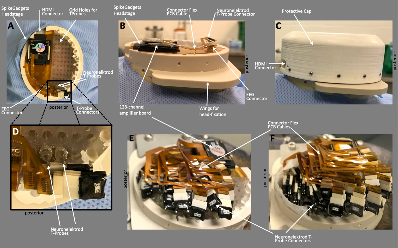
Top-down (A) and lateral view (B&C) of the MePhys platform on the animal’s head. The platform is designed around the 1024 channel modular headstage from SpikeGadgets that is located over the left hemisphere. (C) A mini-HDMI connector allows us to stream data without removing the protective cap. Thus, the protective cap can remain in place for months and only needs to be removed for maintenance. (A) A regular grid of 72 holes covers the right hemisphere. (A-D) The MePhys platform after the first two electrode shafts have been implanted over occipital cortex. The platform after the third (E) and final (F) surgery is almost completely covered by the Plexon T-Probe connectors and the flex-PCB cables that connect the electrodes to the headstage
Top-down (A) and lateral view (B&C) of the MePhys platform on the animal’s head. The platform is designed around the 1024 channel modular headstage from SpikeGadgets that is located over the left hemisphere. (C) A mini-HDMI connector allows us to stream data without removing the protective cap. Thus, the protective cap can remain in place for months and only needs to be removed for maintenance. (A) A regular grid of 72 holes covers the right hemisphere. (A-D) The MePhys platform after the first two electrode shafts have been implanted over occipital cortex. The platform after the third (E) and final (F) surgery is almost completely covered by the Plexon T-Probe connectors and the flex-PCB cables that connect the electrodes to the headstage
Top-down (A) and lateral view (B&C) of the MePhys platform on the animal’s head. The platform is designed around the 1024 channel modular headstage from SpikeGadgets that is located over the left hemisphere. (C) A mini-HDMI connector allows us to stream data without removing the protective cap. Thus, the protective cap can remain in place for months and only needs to be removed for maintenance. (A) A regular grid of 72 holes covers the right hemisphere. (A-D) The MePhys platform after the first two electrode shafts have been implanted over occipital cortex. The platform after the third (E) and final (F) surgery is almost completely covered by the Plexon T-Probe connectors and the flex-PCB cables that connect the electrodes to the headstage

(A) Exploded design of the MePhys platform with the main structural and functional elements: crown, spider, wall, grid, baseplate, headstage and cap. (B) Schematic of the electrode – guidepin – grid system. (C) The intracranial contacts (blue dots) of the MePhys prototype are arranged in 14 coronal slices from posterior to anterior in the panels from bottom right to top left. The antero-posterior position of the slices is indicated by horizontal lines in the top-down schematic of the monkey brain. Slices #1 and #2 were omitted, because they do not contain any currently functional electrode contacts.
Sensory Input
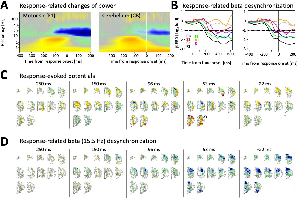
To showcase the ability of MePhys to track hemisphere-wide stimulus-evoked activity, we recorded electric activity from macro- and mesoscopic electrodes in response to short white-noise bursts of varying intensity at three key time-points of the response.
We were able to simultaneously record auditory evoked electric fields from regions in the canonical ascending auditory pathway including brainstem, inferior colliculus, thalamus, and a dense cluster of regions in and around primary auditory cortex. In addition, we also observed auditory evoked responses in the insula; cerebellum; and prefrontal, posterior parietal, and motor cortices.

Motor Output
To probe the ability of MePhys to measure hemisphere-wide motor signals, we used an auditory change detection task during which the animal released a lever when it detected a deviant target tone.
Preprint Highlights
Background Summary
- The temporal resolution of fMRI is limited by the relatively slow hemodynamic response.
- Electrophysiological approaches have either 1) limited spatial resolution (e.g., scalp EEG) or (2) a limited field of view (e.g., microscopic single-cell recordings).
- MePhys yields hemisphere-wide spatial maps of any LFP-based feature, such as sound-evoked electric fields, response-related beta-band desynchronization or resting-state spectra.
- MePhys simultaneously records data from more than 300,000 pairs of electrodes distributed across the entire hemisphere.
- By combining the best features of both EEG and fMRI into a single method in the non-human primate, MePhys will be able to facilitate the comparison between macroscopic findings in humans and microscopic single-cell recordings non-human primates, and thus the translation of findings from rodents, via monkeys to humans, and vice versa.
Top-down (A) and lateral view (B&C) of the MePhys platform on the animal’s head. The platform is designed around the 1024 channel modular headstage from SpikeGadgets that is located over the left hemisphere. (C) A mini-HDMI connector allows us to stream data without removing the protective cap. Thus, the protective cap can remain in place for months and only needs to be removed for maintenance. (A) A regular grid of 72 holes covers the right hemisphere. (A-D) The MePhys platform after the first two electrode shafts have been implanted over occipital cortex. The platform after the third (E) and final (F) surgery is almost completely covered by the Plexon T-Probe connectors and the flex-PCB cables that connect the electrodes to the headstage
Top-down (A) and lateral view (B&C) of the MePhys platform on the animal’s head. The platform is designed around the 1024 channel modular headstage from SpikeGadgets that is located over the left hemisphere. (C) A mini-HDMI connector allows us to stream data without removing the protective cap. Thus, the protective cap can remain in place for months and only needs to be removed for maintenance. (A) A regular grid of 72 holes covers the right hemisphere. (A-D) The MePhys platform after the first two electrode shafts have been implanted over occipital cortex. The platform after the third (E) and final (F) surgery is almost completely covered by the Plexon T-Probe connectors and the flex-PCB cables that connect the electrodes to the headstage
(A) Exploded design of the MePhys platform with the main structural and functional elements: crown, spider, wall, grid, baseplate, headstage and cap. (B) Schematic of the electrode – guidepin – grid system. (C) The intracranial contacts (blue dots) of the MePhys prototype are arranged in 14 coronal slices from posterior to anterior in the panels from bottom right to top left. The antero-posterior position of the slices is indicated by horizontal lines in the top-down schematic of the monkey brain. Slices #1 and #2 were omitted, because they do not contain any currently functional electrode contacts.
Sensory Input
To showcase the ability of MePhys to track hemisphere-wide stimulus-evoked activity, we recorded electric activity from macro- and mesoscopic electrodes in response to short white-noise bursts of varying intensity at three key time-points of the response.
We were able to simultaneously record auditory evoked electric fields from regions in the canonical ascending auditory pathway including brainstem, inferior colliculus, thalamus, and a dense cluster of regions in and around primary auditory cortex. In addition, we also observed auditory evoked responses in the insula; cerebellum; and prefrontal, posterior parietal, and motor cortices.
Motor Output
To probe the ability of MePhys to measure hemisphere-wide motor signals, we used an auditory change detection task during which the animal released a lever when it detected a deviant target tone.