A guide to design the optimal probe for your experiments
Newsletter from August 17th, 2018 which provide a guide for designing your U, V, or S-Probe.


|
Plexon offers customers the ability to design customized probes for electrophysiology research. Because researchers can modify each specification of a probe, the process of creating a probe may seem overwhelming. This email will help you design an optimal probe for your experiment. For a complete list of probe specifications, download our Plexon Probes Data Sheet
V-PROBE >
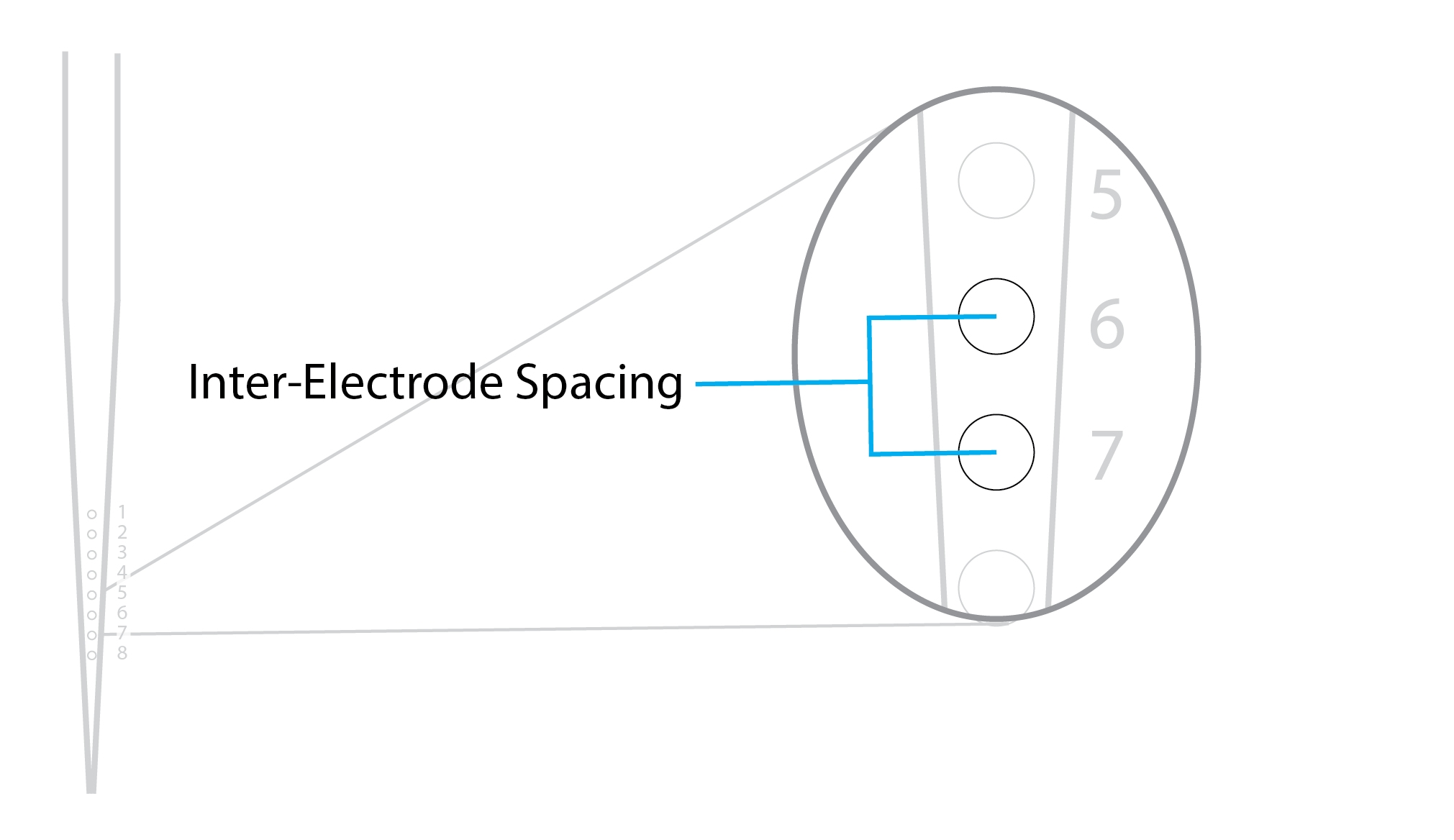
8 QUESTIONS FOR DESIGNING YOUR PROBE1. STUDYING A REGION WITH A HIGH DENSITY OF NEURONS?  Plexon U, V, and S-Probes are available in 8,16, 24, and 32 channel configurations If your experiments involve brain regions with a high density of neurons, such as the amygdala, we suggest using higher channel count probes. We also recommend considering a stereotrode or tetrode configuration. The number of channels you select will influence the diameter of the probe. 2. INTERESTED IN RECORDING FROM SINGLE UNITS? Electrode site diameters range from 15 to 40 microns. Most customer choose 15 microns for resolving single units. Increasing the electrode diameter will increase the diameter of the probe shaft. Recording sites can be 50, 75, 100, 150, or 200 microns apart (inter-electrode spacing):
3. RECORDING FROM DEEP BRAIN STRUCTURES?  Total Probe Length: The length of the probe is measured from tip to connector (Reinforcement Tube + Shank Length). The total length of your probe can be 30 to 150mm. Diameter: The probe diameter will depend on the channel count and depth of target recording site. Adding fluid channels or fiber optics will also influence the diameter. 4. HOW DO I DECIDE ON SPECIFICATIONS FOR MY REINFORCEMENT TUBE?  The reinforcement tube is used to connect the probe to a microdrive. Reinforcement tube length: 5 – 125 millimeters Note: The reinforcement tube must be at least 25mm less than the total probe length. Reinforcement tube diameter is dependent on the probe diameter. 5. HOW CLOSE CAN THE FIRST ELECTRODE SITE BE TO THE TIP OF THE PROBE? 
Distance from tip to first electrode site will be dependent on probe type and probe diameter. If you are looking for a probe that has the smallest possible distance between electrode site and tip, then you may want to consider the V-Probe. Another benefit of the V-Probe is that the distance to first site remains constant regardless of your other configuration options. 6. INTERESTED IN ADDING DRUG-DELIVERY, OR OPTOGENETIC STIMULATION TO YOUR PROBE? Fluid channels and optic fibers can be placed between any channels in a U, or S-Probe; however fluid channels and optic fibers must be placed in the first half of sites closest to connector in a V-Probe. You can add up to 4 fluid /optic channels to your probe:
 7. INTERESTED IN LEARNING ABOUT HOW OTHER RESEARCHERS ARE USING THESE PROBES?  Over 200 labs across the world have ordered and are using these probes. Contact us to learn how researchers are using Plexon probes. 8. INTERESTED IN RECORDING FROM A CHRONIC PROBE? This email primarily offered guidance for the Plexon S, U, and V-Probes. If the below probes are more fitting for your research, please contact a Plexon Sales Engineer.
Thanks for reading! We hope these steps help with your probe-design process. Already have a probe configuration in mind? Start your order with a quote request today. If you enjoyed reading and have friends or colleagues who may be interested in our electrodes, forward this email to them. They can sign up to receive our future newsletters here: plexon.com/newsletter-signup Until next time, – The Plexon Team N-Form and Modular Bionics are trademarks of Modular Bionics Inc. U.S. Patent No. 9,095,267 and patents pending © 2018 All Rights Reserved. Plexon Inc |